What Is Vitreomacular Traction?
Vitreous is the clear, jelly-like substance that fills the middle of your eyes. The vitreous is connected to your retina and macula by millions of tiny fibers.
The macula is responsible for your detailed central vision.
Vitreomacular traction is a condition in which the vitreous of the eye pulls on the macula, which may damage the macula. The vitreous is connected to your retina and macula by millions of tiny fibers.
The vitreous starts to shrink and pulls away from the retina. Eventually, the vitreous will completely detach from the retina. This condition, called posterior vitreous detachment (PVD), is a regular part of aging.
However, sometimes the vitreous doesn’t completely detach, and a portion remains stuck to the macula. When this occurs, the vitreous can pull on the macula, leading to vitreomacular traction. Untreated, vitreomacular traction can lead to macular complications such as holes, scar tissue, and swelling. It can also cause the retina to become detached, a condition called retinal detachment.
What Causes Vitreomacular Traction?
Vitreomacular traction is typically caused by the vitreous not detaching completely from the retina as you age (PVD). Some factors may increase your risk of developing vitreomacular traction, including:
- Age-related macular degeneration (AMD): AMD is a condition common with aging in which the macula begins to deteriorate.
- Diabetic retinopathy and diabetic macular edema: Diabetic retinopathy occurs when diabetes damages the blood vessels in the retina.
- Diabetic retinopathy can lead to proliferative diabetic retinopathy (PDR) — when abnormal blood vessels grow on the retina — and to diabetic macular edema, a condition in which fluid builds up in the macula. These conditions also increase your risk of vitreomacular traction.
- Extreme nearsightedness
- Retinal vein occlusion: In a retinal vein occlusion, one of the veins leading out of the eye becomes blocked
What Are the Symptoms of Vitreomacular Traction?
Vitreomacular traction often leads to changes in vision. These may include:
- Decrease in vision sharpness
- Distortions in vision that make straight lines look wavy (metamorphosis)
- Objects looking smaller than their actual size (micropsia)
- Seeing flashes of light in the eye (photopsia)
These symptoms may come on slowly. They also mimic many other eye conditions, so it’s important to see a doctor if you experience these symptoms.
How Is Vitreomacular Traction Diagnosed?
Your ophthalmologist will need to examine your eye to diagnose vitreomacular traction. There are a few different imaging tests they can use to do this, including:
- Dynamic B-scan ultrasound: a type of ultrasound test that uses sound waves to create an image of the inside of the body. Dynamic B-scan ultrasound can show the doctor the relationship between the vitreous and retina.
- Fluorescein angiography: In fluorescein angiography, your doctor injects yellow dye into your arm. This dye travels to the blood vessels of your eye, and your doctor uses a special camera to take pictures of the eye. The dye shows up brightly on the photos, helping your doctor see where the issues may be.
- Optical Coherence Tomography: Optical coherence tomography is the test most commonly used to diagnose vitreomacular traction. This test uses light waves to produce cross-sectional images of the retina’s layers.
How Is Vitreomacular Traction Treated?
There are generally four treatment options for vitreomacular traction: monitoring, medication, pneumatic vitreolysis, and surgery. Your doctor will recommend a treatment based on the severity of your vitreomacular traction.
Vitrectomy Surgery
In severe cases, your ophthalmologist may decide to perform a type of surgery called a vitrectomy.
A vitrectomy must be done in a surgery center. Your doctor will make a small cut in your eye and use a microscope to examine the inside of your eye. They then use minimal tools to sever the connection between the vitreous and retina and repair any damage to the retina.
Monitoring
In some cases, vitreomacular traction is mild enough not to cause vision changes. In this case, your ophthalmologist will likely schedule follow-up visits to monitor your condition. They may also ask you to monitor your vision yourself with an Amsler grid.
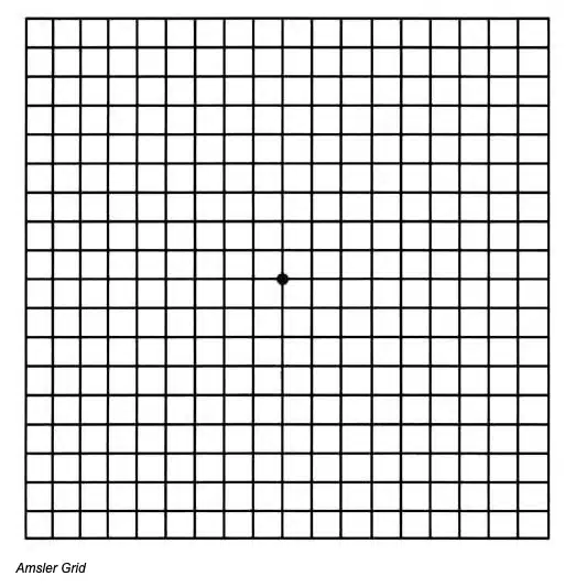
An Amsler grid is a square grid of straight black lines with a dot in the center. To use an Amsler grid¹:
- Hold the grid about a foot away from your face in good light. Wear whatever glasses or contact lenses you usually use.
- Cover the unaffected eye.
- With the uncovered eye, focus on the center dot.
- If the grid starts to become distorted — for example, if the lines become wavy or areas look blurry or blank — that’s a sign that there are problems with your vision.²
- If you are dealing with vitreomacular traction in both eyes, repeat the test on the other eye.
1 Amsler Grid eye test
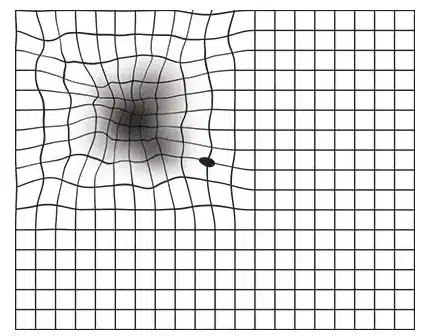
2 What someone with AMD can see. Wavey lines and black spots.
Sometimes, mild cases of vitreomacular traction resolve on their own without treatment.
Medication
A medication called ocriplasmin has proven to be a good option for people with vitreomacular traction. It works by dissolving the fibers that keep the vitreous stuck to the macula. A single injection into the center of the eye is given with Ocriplasmin.
Pneumatic vitreolysis
Pneumatic vitreolysis is a procedure where your doctor injects a small gas bubble into your eye. The goal is to break the bond between the vitreous and the macula. To make that work, you will need to look down several times an hour for a few days to get the bubble into the correct position.
Sometimes, your doctor will use pneumatic vitreolysis in combination with medication to encourage the vitreous to separate from the macula fully.
Get Help Today
The team at M&M Eye Institute is here to help restore your vision. Each of our eye centers uses cutting-edge techniques and advanced technology to treat patients. If vision problems are plaguing you, contact M&M Eye Institute today.